Comprehensive Guide to CT Scan for Lung Cancer: Essential Insights for Accurate Diagnosis and Effective Treatment

In the realm of modern medicine, especially within Health & Medical, Sports Medicine, and Physical Therapy sectors, technological advancements have revolutionized how clinicians diagnose and treat complex conditions. Among these innovations, the CT scan for lung cancer stands out as a cornerstone imaging modality that significantly influences patient outcomes through early detection and precise characterization of lung anomalies.
Understanding the Role of CT Scans in Lung Cancer Diagnosis
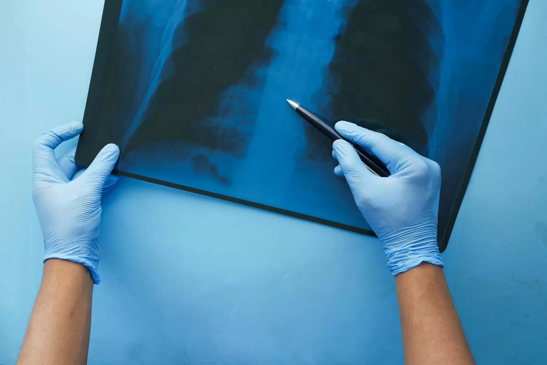
Computed Tomography (CT) scans have transformed pulmonary medicine by providing high-resolution, cross-sectional images of the lungs and surrounding structures. When it comes to lung cancer, the ability to visualize small nodules and differentiate benign from malignant masses is crucial. The CT scan for lung cancer is often the first step in identifying suspicious lesions, guiding biopsy procedures, and staging the disease to plan appropriate interventions.
Why is the CT scan for lung cancer So Critical?
The importance of a CT scan for lung cancer lies in several key functions:
- Early Detection: Detects small nodules that are often invisible on standard X-rays, increasing the chance of diagnosing lung cancer at an early, more treatable stage.
- Differentiation of Lesions: Helps distinguish malignant tumors from benign growths, infections, or scars.
- Staging Accuracy: Determines the extent of disease spread within the chest and beyond, vital for selecting suitable treatment options.
- Guidance for Biopsy: Assists in precisely targeting lesions for minimally invasive biopsies, improving diagnostic accuracy.
- Monitoring Response: Tracks the effectiveness of treatments over time, informing adjustments in therapy.
How the CT Scan for Lung Cancer Is Performed
The procedure of a CT scan for lung cancer is minimally invasive and generally quick, typically taking between 10 to 30 minutes. Patients are required to follow specific instructions to ensure clear images:
- Preparation: Patients may need to fast for several hours prior to the scan if contrast dye is used.
- Positioning: Lying flat on the scanner table, usually face up, with arms raised above the head.
- Contrast Administration: An intravenous contrast dye may be administered to enhance imaging quality and detail.
- Scanning: The scanner rotates around the chest, capturing multiple cross-sectional images.
- Post-Procedure: Patients may be observed briefly for any adverse reactions to contrast media, and instructions for aftercare are provided.
Advancements in CT Technology Enhancing Lung Cancer Detection
The evolution of CT technology has dramatically increased the sensitivity and specificity of lung cancer detection. Notable advancements include:
- Low-Dose CT (LDCT): Reduces radiation exposure while maintaining diagnostic accuracy, making screening more accessible and safer for high-risk populations.
- High-Resolution CT (HRCT): Provides detailed imagery of lung tissue, instrumental in characterizing small nodules.
- Artificial Intelligence and Computer-Aided Detection (CAD): Assists radiologists by highlighting suspicious areas, improving detection rates, and reducing human error.
- 3D Imaging and Reconstruction: Allows comprehensive spatial visualization of lung lesions, aiding in surgical planning and treatment evaluation.
The Importance of Regular Screening with CT for High-Risk Patients
National guidelines emphasize the significance of routine screening, particularly for individuals with heavy smoking histories, age over 55, or other risk factors. Low-dose CT screening programs have demonstrated a reduction in lung cancer mortality by enabling earlier diagnosis. Regular screening using CT scans for lung cancer can detect tumors before symptoms appear, vastly improving treatment success rates and survival outcomes.
Risks and Limitations of CT Scans in Lung Cancer Detection
While CT scans are highly valuable, it is essential to recognize their limitations and potential risks:
- Radiation Exposure: Despite low-dose protocols, repeated scans contribute to cumulative radiation doses.
- False Positives: Can lead to unnecessary invasive procedures or anxiety.
- Overdiagnosis: Detecting indolent tumors that might not require treatment, potentially leading to overtreatment.
- Contrast Reactions: Allergic responses or kidney issues may occur in some patients receiving contrast dye.
Complementary Diagnostic Tools to the CT Scan for Lung Cancer
Though a CT scan for lung cancer is critical, it is often used in conjunction with other diagnostic approaches to confirm findings and improve accuracy:
- Positron Emission Tomography (PET): Combines metabolic activity imaging to distinguish benign from malignant nodules.
- Biopsy Procedures: Such as transthoracic needle biopsy or bronchoscopy to obtain tissue samples for histopathology.
- Blood Tests: Research is ongoing into tumor markers that may support diagnosis or monitor disease progression.
- Genetic Testing: Identifies mutations for targeted therapies, enhancing personalized treatment plans.
Empowering Patients with Knowledge and Support
Understanding the importance of CT scan for lung cancer empowers patients to seek timely screening and follow-up care. Early detection through advanced imaging can be life-saving. Patients should discuss their risk factors with healthcare professionals, especially those in Health & Medical, Sports Medicine, and Physical Therapy settings, to develop personalized screening strategies.
Choosing the Right Facility for CT Lung Cancer Screening
When considering a CT scan for lung cancer, it is vital to select a reputable, accredited imaging center with state-of-the-art equipment and experienced radiologists. Look for centers that follow established guidelines for screening and diagnostic protocols. For residents in Singapore, Hellophysio.sg offers comprehensive lung health assessments utilizing the latest CT technology, ensuring accuracy and safety in diagnosis.
Conclusion: The Future of Lung Cancer Detection Using CT Imaging
The landscape of lung cancer diagnosis continues to evolve with technological innovations in CT imaging. The CT scan for lung cancer remains an indispensable tool, enabling early detection, guiding precise treatments, and ultimately saving lives. As research advances, NEW modalities, including AI-assisted analysis and integrated diagnostic approaches, promise to further enhance outcomes and reduce mortality rates.
Incorporating routine CT screening programs, fostering awareness, and ensuring access to quality facilities will pave the way for a healthier future. Staying informed and proactive about lung health is paramount—embrace the benefits of advanced imaging to detect lung cancer early and improve your chances for successful treatment.


